
Definition Of X Ray Pdf defitioni
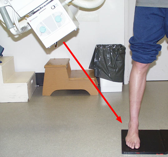
Volume 2 No. 1 Positioning: Recommended Beam Centers Center the x-ray beam directly over the area of interest. Visualize how the image would look on a monitor. Move the patient and position the area of interest along the long axis of your collimated field, rather than rotating the collimator.
Ballinger Radiographic Positioning Pdf Free
Go to: Definition/Introduction Imaging of the body is often complicated by the fact that anatomic structures overlap each other. Diagnostic accuracy of radiographs generally refers to how well an exam can predict the presence (or absence) of a disease or condition.
XRay Positioning Guide Toes Medical Professionals
About this app. -Position of the patient. -Chassis to use. -Focus focus film. -Director ray. -Utility. -QA. In addition you will be able to visualize examples of radiographs and the positioning of the patient, which will make your study much more visual and enjoyable. Study in a different, more intuitive and interactive way.

X Ray Positioning Chart With Images Pdf
The iRadTech app is a radiographic positioning guide for Apple and Android smartphones and tablets for $24.99 in the app stores. Watch a video of iRadTech in action It is also available as a web app, delivered to a browser, so that it is platform independent. See below for further information. Similar to x-ray pocket guide or reference booklet
X Ray Positioning Chart Free Download evermagic
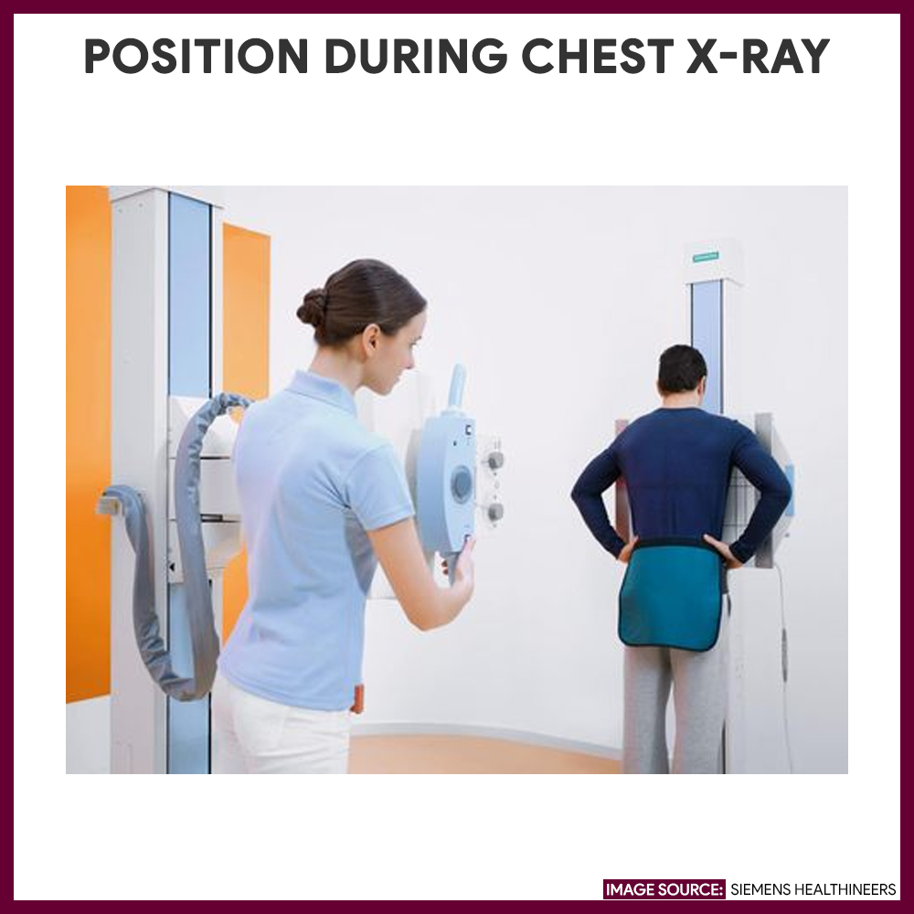
AP, PA, Lateral Anterior-Posterior (AP) radiographs are taken with the patient facing the x-ray tube, so that the x-ray beam enters their anterior side, and exits posteriorly. Posterior-Anterior (PA) films are performed while the patient faces away from the x-ray tube. The x-ray beam goes in their posterior and comes out their anterior.
Radiographic Positioning Radiology Key
A standard Radiography technique chart is a written table that contains the following technical data to help radiographers obtain a consistent, standardized image while using the lowest radiation dose possible: The body part imaged (hand, foot, skull, etc) The kV or kilovolts required for the image (how strong of a beam)
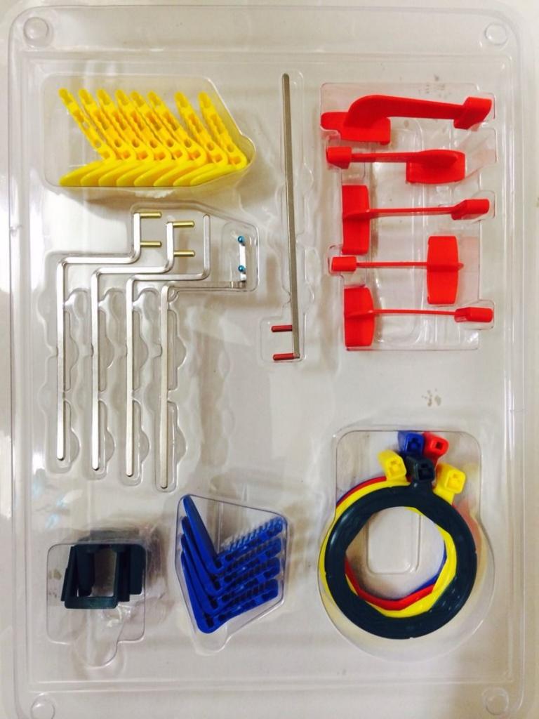
XRay Positioning System MR Dental
USING THE CHARTS This chapter is designed as a quick reference guide to radiographic positioning and technique. Technical tips and supplemental views are provided to aid in obtaining optimal film quality using the most appropriate views.
เครื่องมือทันตกรรม X Ray Positioning Xcp 3000 jodyaccessories.th ThaiPick
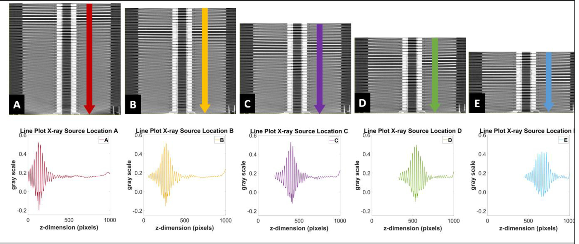
It is recommended that purchasing digital X-ray equipment with high detective quantum efficiency detectors, and then optimising the exposure chart for use with these detectors is of high.
เครื่องมือทันตกรรม X Ray Positioning Xcp 3000 jodyaccessories.th ThaiPick
Tips Take at least two views of each anatomic region—remember, you're capturing a three-dimensional object. Center the x-ray beam directly over the area of interest. Visualize how the image would look on a monitor. Move the patient and position the area of interest along the long axis of your collimated field, rather than rotating the collimator.
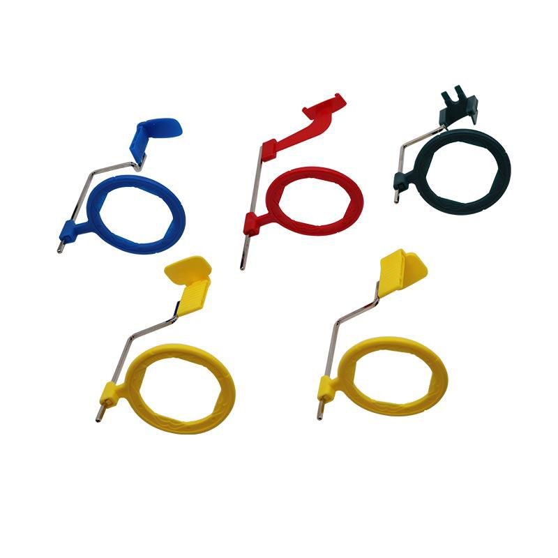
X Ray Positioning System Mayfair Dental Supplies
"The X-Ray Lady" 6511 Glenridge Park Place, Suite 6 Louisville, KY 40222 Telephone (502) 425-0651 Fax (502) 327-7921 Web address www.x-raylady.com Email address [email protected] Review of Radiographic Anatomy & Positioning and Pediatric Positioning Approved for 5 Category A Credits American Society of Radiologic Technologists (ASRT)

DENTAL XRAY Positioning System Shanghai Dental Material
Download full-text PDF Read full-text.. Figure 8 is a flow chart for positioning training.. It is desirable to perform the positioning accurately. As X-ray films are usually used in.
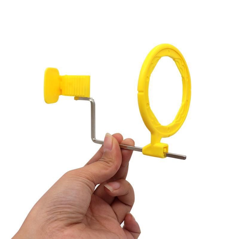
X Ray Positioning System Mayfair Dental Supplies
Position the opposite limb out of the way by taping around the carpus and pulling it across the body in a caudodorsal direction, and attach the tape to the edge of the table. Pull the affected limb cranially and position it in a normal walking motion, using tape or a sandbag to secure it in place (FIGURE 22).
เครื่องมือทันตกรรม X Ray Positioning Xcp 3000 jodyaccessories.th ThaiPick
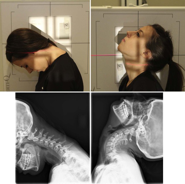
erect: either standing or sitting. decubitus : lying down. supine : lying on back. Trendelenburg position: the patient is supine (on an inclined radiographic table) with the head lower than the feet. prone : lying face-down. lateral: side touches the cassette. right lateral: right side touches the cassette. left lateral: left side touches the.
Podiatry Xray Positioning wikiRadiography
Download Free PDF. Clark's Positioning in Radiography, 12th ed, Arnold. Clark's Positioning in Radiography, 12th ed, Arnold.. Standing position and patient factors should be considered when defining "optimal" acetabular orientation. Download Free PDF View PDF. Acta Orthopaedica.

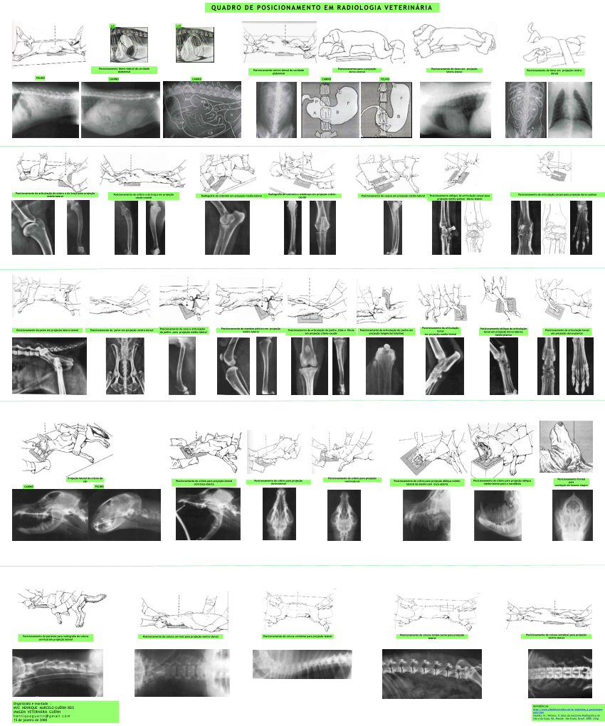
socialpetworking Vet medicine, Veterinary radiology, Vet student
With more than 400 projections, Merrill's Atlas of Radiographic Positioning & Procedures, 14th Edition makes it easier to for you to learn anatomy, properly position the patient, set exposures, and take high-quality radiographs. This definitive text has been reorganized to align with the ASRT curriculum — helping you develop the skills to produce clear radiographic images.
X ray positioning pictures labquiz
Click on the Download Button below to start downloading Fujifilm X-ray positioning chart PDF . It's a direct download link. It will instantly start downloading the pdf. Tags Fujifilm X-ray positioning chart: Newer KUB MCQs (Kidneys, Ureters, and Bladder) For Govt Exam Practice | SSC, UPSC | Bsc Radiology, BXRT Older