Bacterial Cell Structure and Function
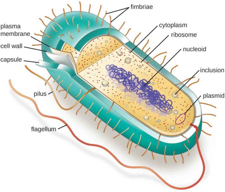
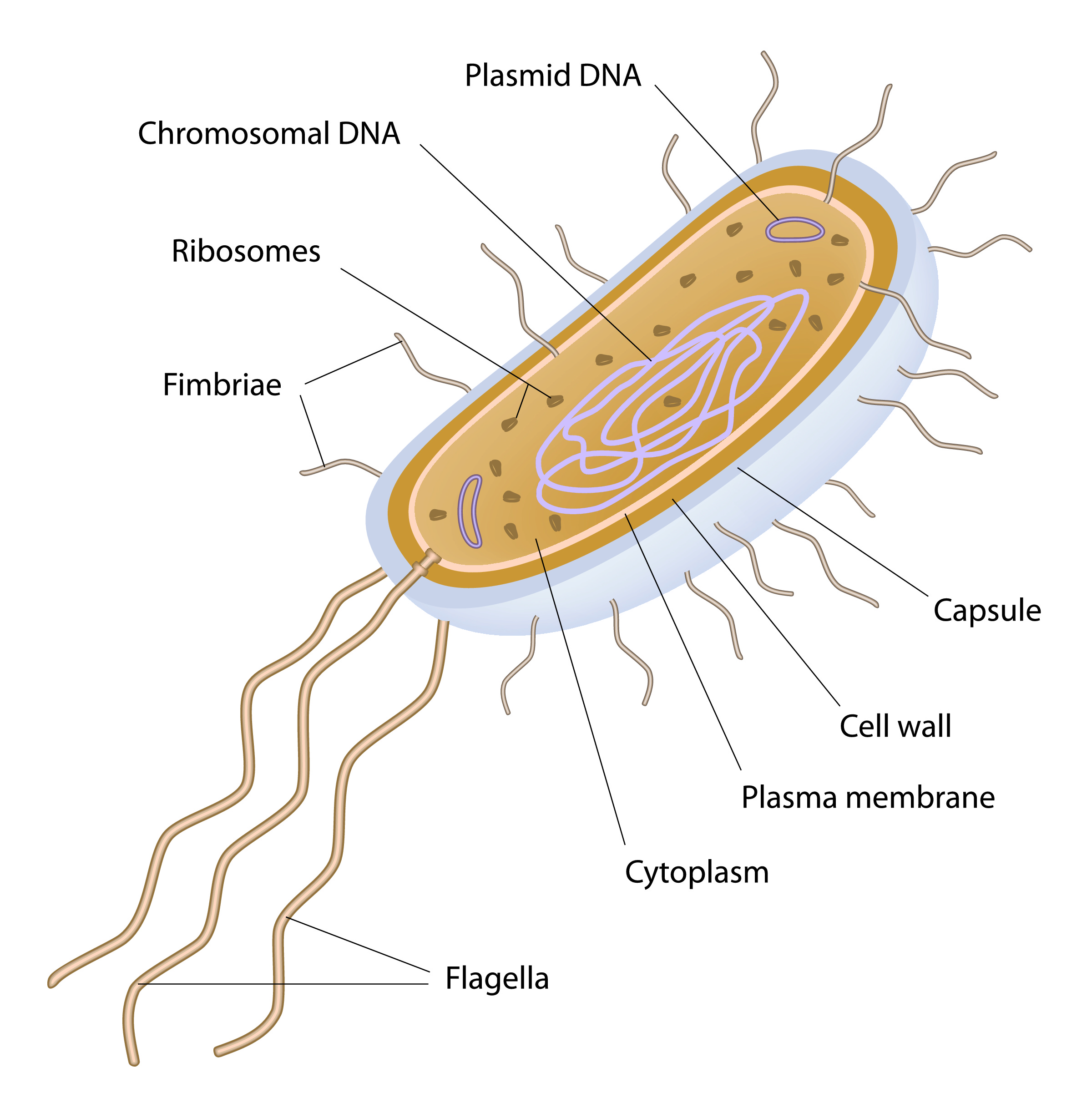
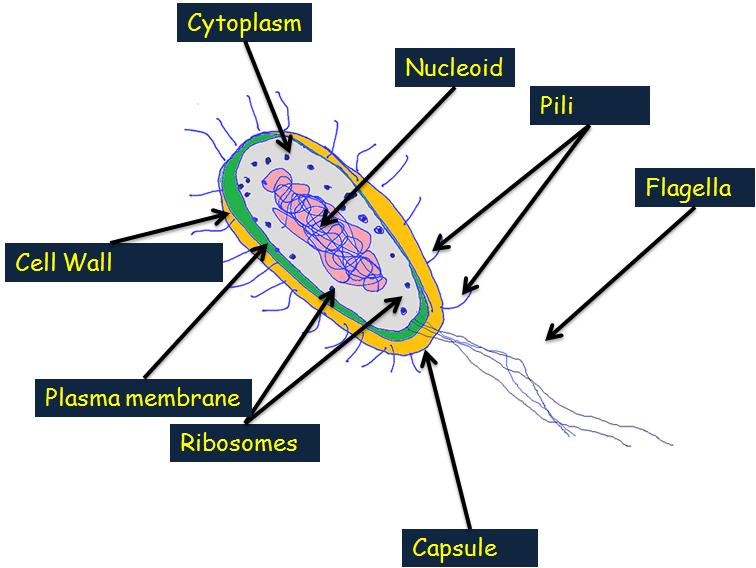
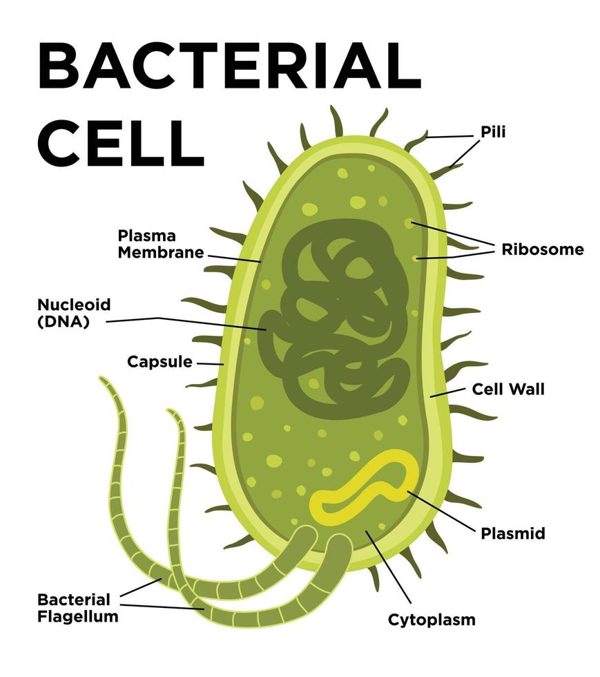
Bacteria Diagram with Labels Bacterial cells have simpler internal structures like Pilus (plural Pili), Cytoplasm, Ribosomes, Capsule, Cell Wall, Plasma membrane, Plasmid, Nucleoid, Flagellum, etc. Labeled Bacteria diagram Eukaryotes have been shown to be more recently evolved than prokaryotic microorganisms.

Picture Prokaryotic cell, Cell structure, Bacterial cell structure
Key points: Prokaryotes are single-celled organisms belonging to the domains Bacteria and Archaea. Prokaryotic cells are much smaller than eukaryotic cells, have no nucleus, and lack organelles. All prokaryotic cells are encased by a cell wall. Many also have a capsule or slime layer made of polysaccharide.
Eukaryotes and Prokaryotes worksheet from EdPlace
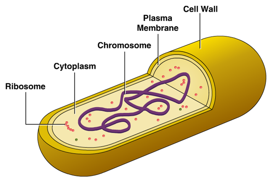
In this article we will discuss about the cell structure of bacteria with the help of diagrams. A bacterial cell (Fig. 2.5) shows a typical prokaryotic structure. The cytoplasm is enclosed by three layers, the outermost slime or capsule, the middle cell wall and inner cell membrane.
Bacteria stock vector. Illustration of bacteria, ribosome 41152551
coccus (circle or spherical) bacillus (rod-like) coccobacillus (between a sphere and a rod) spiral (corkscrew-like) filamentous (elongated) Cell shape is generally characteristic of a given bacterial species, but can vary depending on growth conditions.

Label the Bacterial Cell Key New Unit 1 Cells and Cell Processes Ppt Cell processes, Cell wall
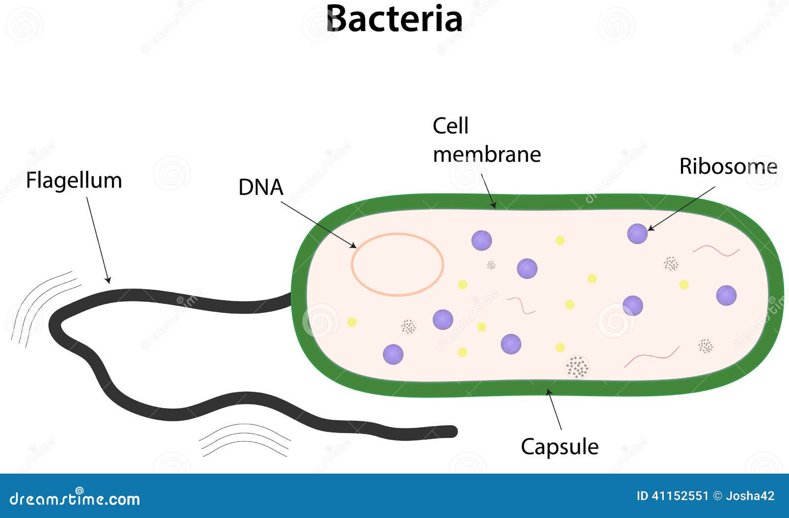
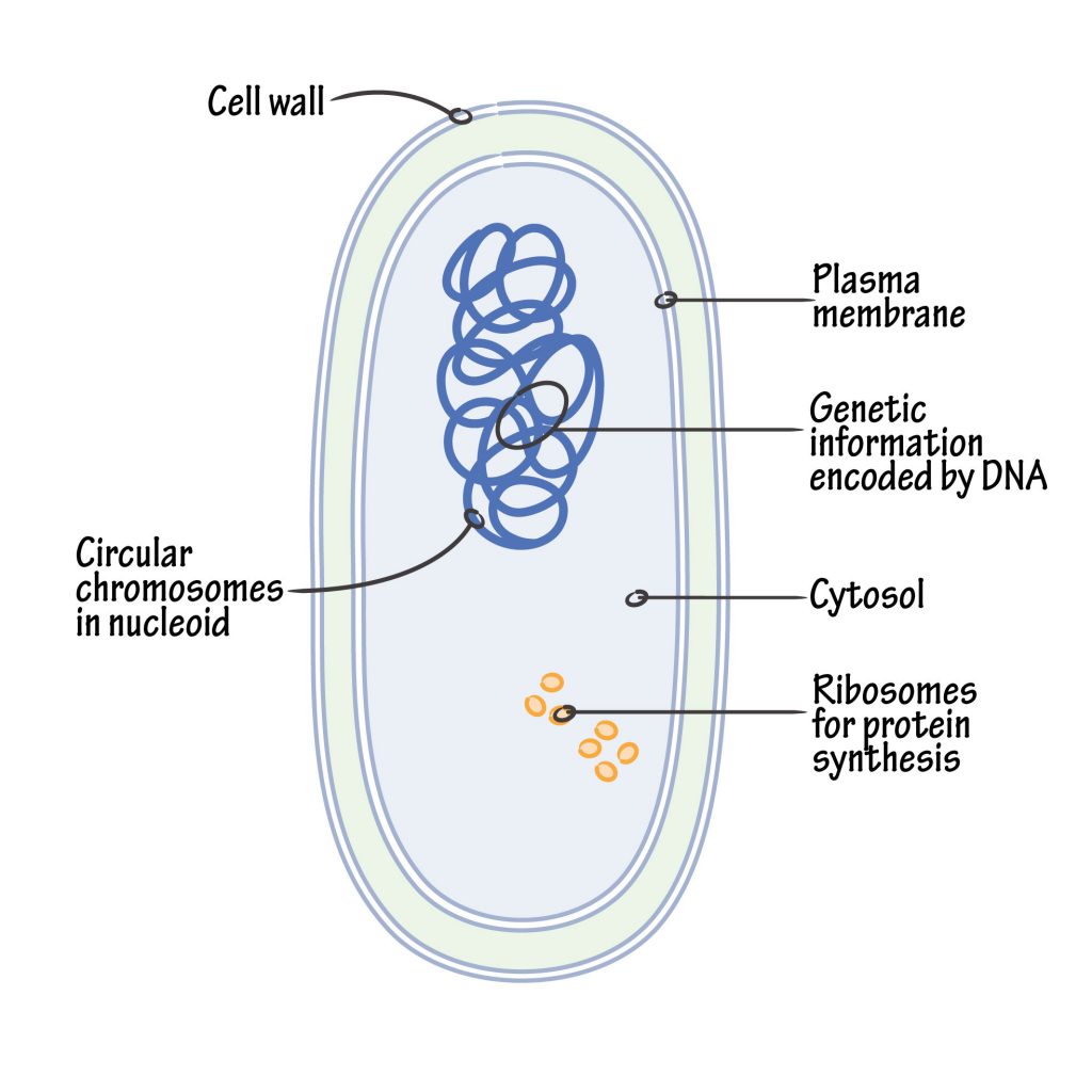
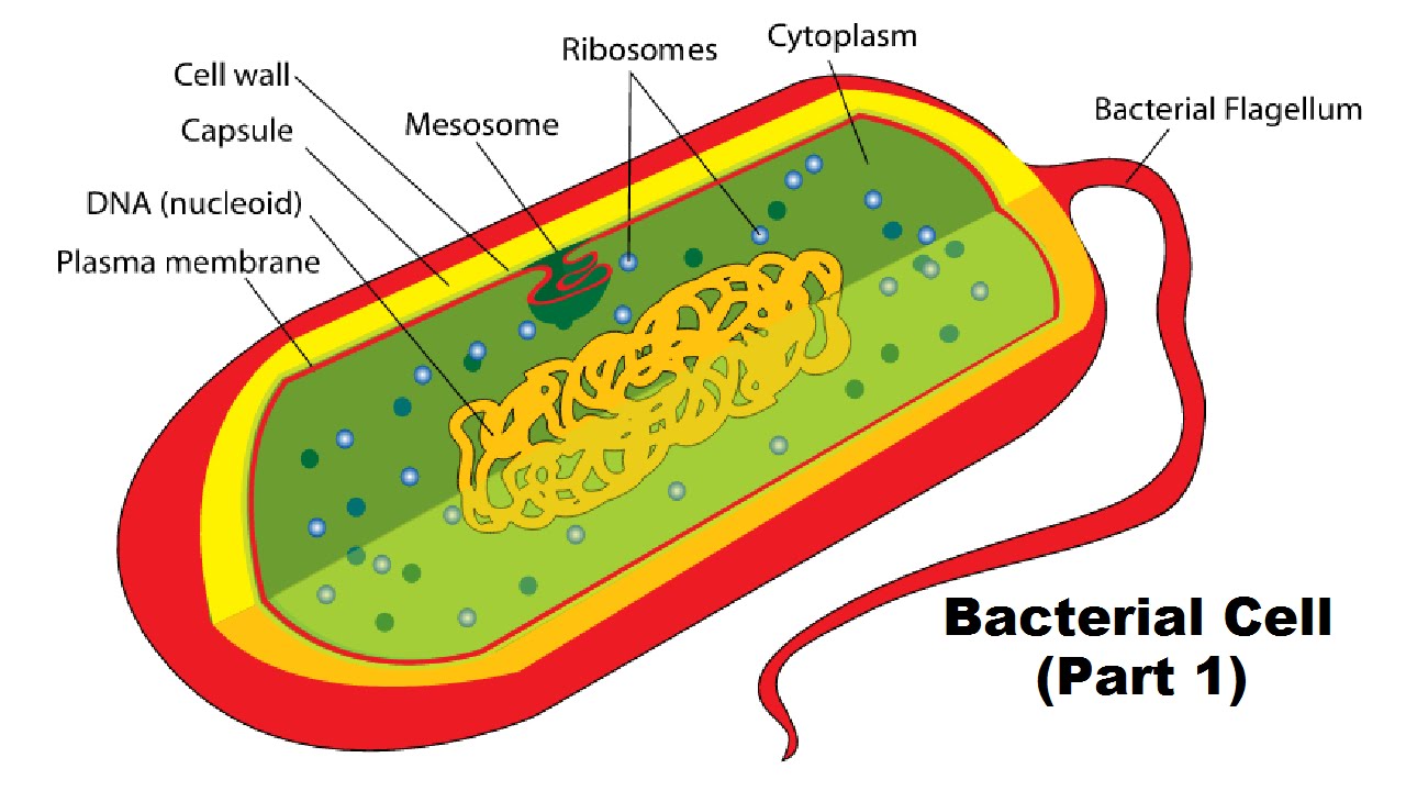
Bacterial cell have simpler internal structure. It lacks all membrane bound cell organelles such as mitochondria, lysosome, golgi, endoplasmic reticulum, chloroplast, peroxisome, glyoxysome, and true vacuole. Bacteria also lacks true membrane bound nucleus and nucleolus. The bacterial nucleus is known as nucleoid.

Structure and Function of Prokaryotic Cells
Bacteria Diagram representing the Structure of Bacteria Ultrastructure of a Bacteria Cell The structure of bacteria is known for its simple body design. Bacteria are single-celled microorganisms with the absence of the nucleus and other c ell organelles; hence, they are classified as prokaryotic organisms.

Bacterial Cell Diagrams 101 Diagrams
The components are: 1. Cell Envelope 2. Cytoplasm 3. Nucleoid 4. Plasmids 5. Inclusion Bodies 6. Flagella 7. Pili and Fimbriae. Bacterial Cell: Component # 1. Cell Envelope: It is the outer covering of protoplasm of bacterial cell. Cell envelope consists of 3 components— glycocalyx, cell wall and cell membrane. (i) Glycocalyx (Mucilage Sheath):
Bacterial Structure Plantlet
Wall-Less Forms: Two groups of bacteria devoid of cell wall peptidoglycans are the Mycoplasma species, which possess a surface membrane structure, and the L-forms that arise from either Gram-positive or Gram-negative bacterial cells that have lost their ability to produce the peptidoglycan structures. Cytoplasmic Structures

Bacterial cell structure Year 12 Human Biology
In this video, we show you how to draw and label a basic bacterial cell. Check out http://eacharya.tumblr.com for more!
Bacterial Cell Labelled Diagram ClipArt Best
August 14, 2021. Bacteria are unicellular. Their structure is a very simple type. Bacteria are prokaryotes because they do not have a well-formed nucleus. A typical bacterial cell is structurally very similar to a plant cell. The cell structure of a bacterial cell consists of a complex membrane and membrane-bound protoplast.
Cellular Structure of Bacteria ZeroInfections
Bacteria (sing. bacterium) are unicellular prokaryotic microorganisms which divide by binary fission. They do not possess nuclear membrane and the nucleus consists of a single chromosome of circular double-stranded DNA helix (Fig. 1.1). Flagella: ADVERTISEMENTS:
Structure of a Bacterial Cell (Part 1) YouTube
Shape and Arrangement-1 Cocci (s., coccus) - spheres diplococci (s., diplococcus) - pairs streptococci - chains staphylococci - grape-like clusters tetrads - 4 cocci in a square sarcinae - cubic configuration of 8 cocci Shape and Arrangement-2 bacilli (s., bacillus) - rods coccobacilli - very short rods

Page 1
The schematic diagram of bacterial cell structure is shown in the Fig.1.The bacteria possess the morphological structures for the purpose of performing some physiological functions, e.g. flagella.
Bacterial cell anatomy in flat style. Vector modern illustration. Labeling structures on a
These can rotate or move in a whip-like motion to move the bacterium. Plant and bacterial cell walls provide structure and protection. Only plant cell walls are made from cellulose. The DNA of.

Bacterial Cell Diagrams 101 Diagrams
Bacterium Cell Anatomy Activity Key 1. Flagellum 2. Capsule 3. Cell wall 4. Cell membrane 5. Cytosol 6. Ribosome 7. Pili 8. Plasmid 9. Nucleoid (DNA) Title: Bacterial Cell Coloring Page Author: Ask A Biologist Subject: Bacteria Cell Parts Keywords: Bacteria cell anatomy, cells, bacterium

Bacterial Cell Diagrams 101 Diagrams
A prokaryote is a simple, single-celled organism that lacks a nucleus and membrane-bound organelles.
